[Translate to English:] Biofilmformation
Biofilm formation is the most frequent cause of a faulty voice prosthesis!
Contribution by Dr. M. Leonhard of Vienna
As soon as a shunt valve is inserted in the tracheoesophageal fistula, microbial deposits of bacteria and fungi from the mouth and throat, stomach and deep respiratory tracts start to settle on its surface. Starting with the first cellular adhesions and deposited saliva protections, complex multi layered microbial communities develop, each adapting better to its environment than the one before. These biofilms form their own mucous layer to protect them against dehydration, fluctuating pH levels, food shortages and antimicrobial treatments.
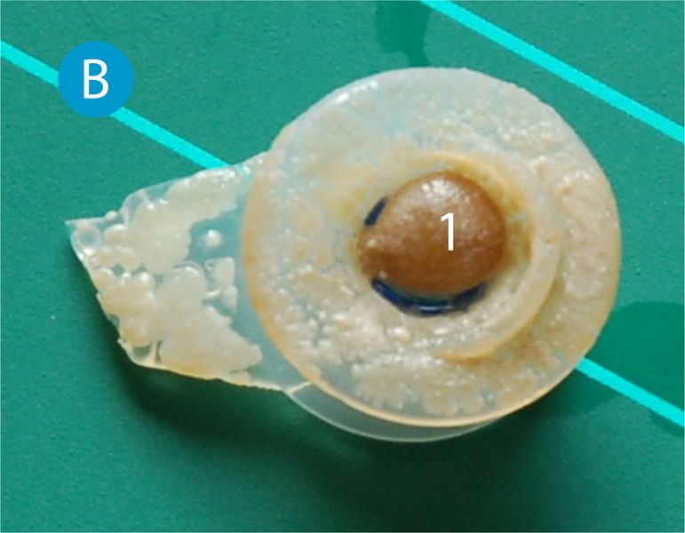
The further consequence is damage to the prosthesis material (fig. B) from ingrowing hyphae, displacement of the prosthesis lumen and blockages of valve parts. The shunt valve starts to leak, and the contents of the food pipe escape through the prosthesis into the wind pipe (aspiration). If this happens, the patient must attend the doctor/SLP in order to be fitted with a new voice valve.
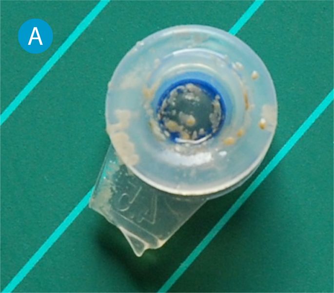
The retention time of a voice prosthesis varies, and averages between 90 and 120 days. The infectious agents most frequently found on shunt valves include the Candida species (C. albicans, C. glabrata, C. crusei, C. tropicalis), Staphylococci (S. aureus), Streptococci (S. parasanuinis), Enterococci and species from the oral cavity. As yet there is no proof of an association between the microbial spectrum and the patient’s diet.
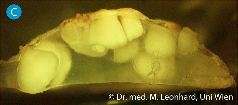
Shunt valves examined under microscope revealed the first signs of material damage in the form of ingrowing fungal hyphae (ill. C), followed by hollowing under the plastic surface, after approx. 3 weeks. This damage results in deformation, discoloration and embrittlement of the prosthesis material, and may cause the valve mechanism to fail. The search for suitable biofilm-resistant materials and valve constructions is currently underway.Biofilms like to settle on the surfaces of prostheses that are situated against the food pipe, starting at surface irregularities, recesses and parts of the valve mechanisms. The construction of a pure biofilm (ill. D) reveals a mucous layer containing a network of water channels (biofilm matrix made of extra-cellular polysaccharides) with various microbial species (fungi and bacteria) embedded in them. As the distance from the biofilm surface increases, the living conditions change for the organisms that have settled there. The available food and oxygen offer decreases; there are fewer pH and moisture fluctuations, and the cells reveal a reduction in metabolic activity. This means that antimicrobial medication has a reduced effect on cells that are deeper down. After a period of maturation, the number of cells stops increasing. Certain enzymes are produced that destabilize the biofilm matrix, meaning that parts of the biofilm tear off and are swept away by the liquid stream (saliva flow). Cleared surfaces are re-colonized.

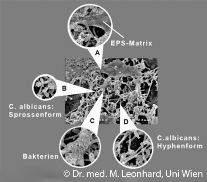
Biofilm structures displayed by electron microscopy
A: Extrapolysaccharide matrix (EPS): mucous layer formed by microorganisms
B+D: typical growth forms of candida fungi: spherical shoots (B) and elongated hyphae (D)
C: bacteria, tiny rods smaller than fungal hyphae in the size comparison